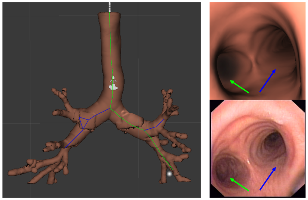
Researchers at the Center for Computer Vision and Bellvitge Hospital – IDIBELL have developed a technology that allows physicians to locate and move around the patients’ bronchi following a planned and localized route thanks to augmented reality techniques, video games and 3D modeling.
The objective of this collaboration is to improve the existing technology in bronchoscopy without adding new instruments during the procedure, as well as to make interventions quicker, more effective and less expensive when obtaining essential biopsies in the early detection of lung cancer .
he team consists of specialists in artificial intelligence and doctors led by Dr. Debora Gil, mathematician and researcher of the Computer Vision Center and professor of the Autonomous University of Barcelona, and Dr. Antoni Rosell, pulmonologist at Bellvitge Hospital and researcher at IDIBELL (Barcelona).
This technology acts as a lung GP; it allows the pneumologists to plan the route to the possible cancer and even to choose what type of technique is going to be used to obtain a biopsy: a clamp, a brush or a needle. With the help of computer vision and 3D modeling techniques, an exact virtual replica of the patient’s lung is created. This replica is introduced in software that, with graphic video game techniques, emulates a bronchoscope navigating the patient’s trachea and bronchus to the marked target and then chooses which diagnostic instrument will finally be used. This ‘video game’ simile, which acts as a localization map, can not only facilitate and reduce the bronchoscopy process, but also reduce drastic interventions with failed results, avoiding repeated interventions for the patient.
This technology also has other applications. It allows accurate measurement of different parts of the bronchial system and can design customized prostheses to the millimeter, with the advantages that this entails. Finally, the associated software makes a reliable calculation of tracheal stenosis (abnormally reduced light) in a record time of just a few seconds. Until now, this diagnosis was made visually, based on the experience of the assigned physician. In this way, researchers have found a diagnosis that moves away from the subjectivity and provides an objective and precise tool.

